Leg Bone Diagram / Uncategorized | Page 2. Most of the leg skeleton has bony prominences and margins that can be palpated and some serve as anatomical landmarks that define the extent of the leg. The ball of the hip joint is made out of the head of the femur, and the femoral head slots into the acetabulum. Beside that, we also come with more related ideas as follows free printable human anatomy coloring pages, lower leg muscle diagram blank and lower limb bones unlabeled. Blank leg bones diagram : It is also known as the calf bone, as it.
ads/bitcoin1.txt
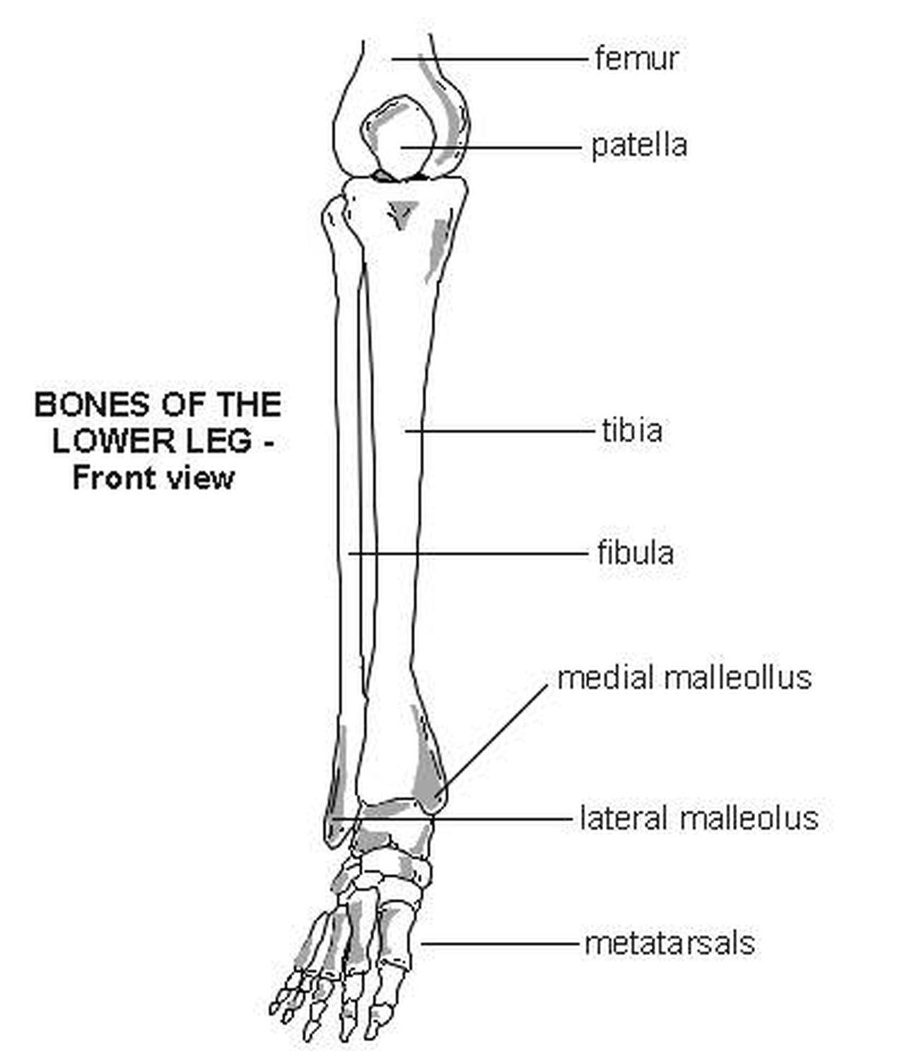
Muscle and tendon pain in legs, muscles and tendons of the leg and foot, muscles and tendons of the lower leg, muscles ligaments and tendons of the lower leg, muscles tendons and ligaments of the upper leg, human muscles, muscle and tendon pain in legs, muscles and tendons of the leg and foot,. Leg bone anatomy diagram diagram of human leg human anatomy diagram 10 / 10 ( 1 vote ) in this image, you will find femur, medial epicondyle of the femur, patella, tibial tuberosity, anterior rest of the tibia, a medial surface of the tibia, lateral epicondyle of the femur, head of the fibula, fibula, medial malleolus of the tibia, lateral. The hip joint gives the leg an incredible range of motion while still providing support to the body's weight. These are the femur, patella, tibia, fibula, tarsal bones, metatarsal bones, and phalanges (see figure 6.51). Start studying lab test 2:
Pictures Of Bones Of The Lower Extremities from healthiack.com The ball of the hip joint is made out of the head of the femur, and the femoral head slots into the acetabulum. There are in all 7 bones, which fall under tarsal bones category. A long bone has a shaft and 2 ends. Beside that, we also come with more related ideas as follows free printable human anatomy coloring pages, lower leg muscle diagram blank and lower limb bones unlabeled. This long bone connects with the knee at one end and the ankle at the other. Distal to the ankle is the foot. The ischium is located just behind the pubis bone. The patella (kneecap) is the sesamoid bone in front of the knee.
The ball of the hip joint is made out of the head of the femur, and the femoral head slots into the acetabulum.
ads/bitcoin2.txt
The tibia and fibula are two long bones that run parallel to each other, forming the scaffold of the leg and providing attachment points for many muscles. This long bone connects with the knee at one end and the ankle at the other. Printable human skeleton diagram u2013 labeled unlabeled and. The ischium is located just behind the pubis bone. The diagram of bones in the ankle and foot is given below: Next to the tibia is the fibula, the thinner, weaker bone of the lower leg. Beside that, we also come with more related ideas as follows free printable human anatomy coloring pages, lower leg muscle diagram blank and lower limb bones unlabeled. Anatomical diagrams of the spine and back. Some types of leg pain can be traced to problems in your lower spine. There are two types of cartilaginous joints: Also called the shin bone, the tibia is the longer of the two bones in the lower leg. Leg (anatomy) leg, limb or appendage of an animal, used to support the body, provide locomotion, and, in modified form, assist in capturing and eating prey (as in certain shellfish, spiders, and insects). A long bone is a bone that has greater length than width.
The tarsal bones in the foot are located amongst tibia, metatarsal bones, and fibula. Now let's look at the tibia bone, which is the larger of the two leg bones, located medially. Every skeletal muscle has three main parts: There are in all 7 bones, which fall under tarsal bones category. Bone diagram forehead (frontal bone) nose bones (nasals) cheek bone (zygoma) upper jaw (maxilla) lower jaw (mandible) breast bone (sternum) upper arm bone (humerus) lower arm bone (ulna) thigh bone (femur) collar bone (clavicle) toe bones (phalanges) ankle bones (tarsals) kneecap (patella) shin bone (tibia) calf bone (fibula) foot bones
Bones of the Leg and Foot | Interactive Anatomy Guide from www.innerbody.com The ball of the hip joint is made out of the head of the femur, and the femoral head slots into the acetabulum. The bones of the leg are the femur, tibia, fibula and patella. Most leg pain results from wear and tear, overuse, or injuries in joints or bones or in muscles, ligaments, tendons or other soft tissues. The pubis curves downward and forwards from the ileum. The ilium is the bone at the top of the waist, while the pubis bones are found just below the ilium. The tibia, commonly known as the 'shin bone', is the largest and most medial of the two. In fact, this bone gets its name from a latin word that literally means shinbone. The foot bones shown in this diagram are the talus, navicular, cuneiform, cuboid, metatarsals and calcaneus.
The hip joint gives the leg an incredible range of motion while still providing support to the body's weight.
ads/bitcoin2.txt
There are two types of cartilaginous joints: Leg (anatomy) leg, limb or appendage of an animal, used to support the body, provide locomotion, and, in modified form, assist in capturing and eating prey (as in certain shellfish, spiders, and insects). When you feel your shinbone, this is what you're feeling. The ischium is located just behind the pubis bone. The hip joint gives the leg an incredible range of motion while still providing support to the body's weight. The head of the femur forms the ball and socket hip joint with the acetabulum of the hip bone. Long bones have a thick outside layer of compact bone and an inner medullary cavity containing bone marrow. The patella (kneecap) is the sesamoid bone in front of the knee. To explain the term in layman's language, it is the heel bone in the skeletal system. The knee joint is the largest joint in the body and is primarily a hinge joint, although some sliding and rotation occur. The tarsal bones in the foot are located amongst tibia, metatarsal bones, and fibula. Start studying lab test 2: This long bone connects with the knee at one end and the ankle at the other.
This long bone connects with the knee at one end and the ankle at the other. Distal to the ankle is the foot. A long bone has a shaft and 2 ends. Most of the leg skeleton has bony prominences and margins that can be palpated and some serve as anatomical landmarks that define the extent of the leg. Long bones have a thick outside layer of compact bone and an inner medullary cavity containing bone marrow.
Leg Bones - Medical Art Library from www.medicalartlibrary.com The femur is the single bone of the thigh. The head of the femur forms the ball and socket hip joint with the acetabulum of the hip bone. The hip joint gives the leg an incredible range of motion while still providing support to the body's weight. These bones join together to make up a socket on the outer rim of the pelvis, the acetabulum. 12 photos of the muscles and tendons of the leg. Muscle and tendon pain in legs, muscles and tendons of the leg and foot, muscles and tendons of the lower leg, muscles ligaments and tendons of the lower leg, muscles tendons and ligaments of the upper leg, human muscles, muscle and tendon pain in legs, muscles and tendons of the leg and foot,. The patella (kneecap) is the sesamoid bone in front of the knee. At the distal end of the femur, two rounded condyles meet the tibia and fibula bones of the lower leg to form the knee joint.
Most leg pain results from wear and tear, overuse, or injuries in joints or bones or in muscles, ligaments, tendons or other soft tissues.
ads/bitcoin2.txt
There are two types of cartilaginous joints: In fact, this bone gets its name from a latin word that literally means shinbone. All the images are in vector format, allowing an optimal web display with zoom and shifting of the anatomical images. It is also known as the calf bone, as it. Distal to the ankle is the foot. 12 photos of the muscles and tendons of the leg. Look at links below to get more options for getting and using clip art. Learn vocabulary, terms, and more with flashcards, games, and other study tools. The diagram of bones in the ankle and foot is given below: When you feel your shinbone, this is what you're feeling. To explain the term in layman's language, it is the heel bone in the skeletal system. He leg's main function in the human is for locomotion and support of the rest of the body. The tibia, commonly known as the 'shin bone', is the largest and most medial of the two.
ads/bitcoin3.txt
ads/bitcoin4.txt
ads/bitcoin5.txt
0 Response to "Leg Bone Diagram / Uncategorized | Page 2"


0 Response to "Leg Bone Diagram / Uncategorized | Page 2"
Post a Comment